How Does Genetic Testing Work?
From a Drop of Blood to a Complete Answer
Authors: Sameer Malik, Sana
The Question Written in Every Cell
For many families, a rare genetic disease begins with years of uncertainty. Seven years is the average time it takes for a family navigating a rare genetic disease to receive a correct diagnosis. A child may miss milestones, tests may come back inconclusive, and one specialist visit may lead to another without a clear answer. In many cases, the clue is already present in the body from birth. It is written in DNA, the instruction system that is present inside our cells.
Genetic testing is the process of reading parts of that DNA to look for changes that may explain a medical condition. Today it is used in newborn screening, cancer care, reproductive medicine, rare disease diagnosis, and many other areas. But the test is not just a blood draw and a result. It is a step-by-step process: collect a good sample, read the DNA accurately, compare it with what is expected, and have experts decide what the findings actually mean.
The Language of Life: DNA, Genes, and the Human Genome
Before we can understand genetic testing, we need to understand what is being tested. The short answer is DNA, or deoxyribonucleic acid. DNA is the instruction material found inside almost every cell in the body.
DNA is made from four chemical letters called nucleotide: A, T, G, and C. These letters are arranged in a very long order. The full human genome has about 3.2 billion pairs of these letters. If the whole sequence were printed in books, it would fill 200+ encyclopedia volumes.
Only some parts of DNA work like direct instructions. These sections are called genes. Humans have about 20,000 genes. Genes sit on chromosomes, which are larger DNA packages. Most people have 46 chromosomes, arranged in 23 pairs, with one chromosome in each pair coming from each parent.
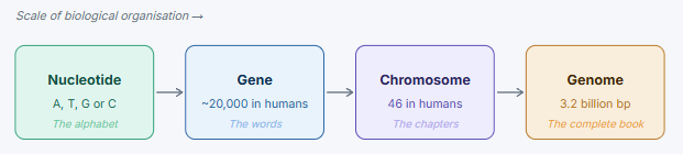
A genetic variant is a change in one or more DNA letters. It is like a spelling difference in the instruction book. Some spelling differences do not matter. Others can change how a gene works.
What is remarkable is that every person shares about 99.9% of their DNA with every other person. The remaining 0.1% is part of what makes each of us different. It also contains the changes that genetic tests look at. Most variants are harmless. A small number can affect how a protein is made, how a gene is switched on or off, or whether an important body process works correctly. The main challenge is finding the few important changes among millions of harmless ones.
What Is Genetic Testing and What It Is Not
Genetic testing looks at DNA or related genetic material to find changes that may matter for health. It can confirm a diagnosis, rule out some conditions, identify carrier status, guide treatment, or show whether a condition can run in a family. It is a medical tool, not a crystal ball. It can only answer questions that current science and the chosen test are able to answer.
This limitation is important. A genetic test produces data, but the data is not useful by itself. The value comes from careful interpretation of it, deciding whether a DNA change actually explains the patient’s symptoms or changes medical care.
A BRIEF HISTORY OF GENETIC TESTING
Karyotyping, which looks at chromosomes under a microscope, became clinically available in the 1950s. It could detect large chromosome changes such as trisomy 21. Sanger sequencing, developed in 1977, made it possible to read individual genes accurately, but only in small sections. Next-generation sequencing, introduced in the mid-2000s, changed the field by allowing many DNA fragments to be read at the same time. Today, a human genome can be sequenced much faster and at far lower cost than during the Human Genome Project.
The Six Stages of Genetic Testing
Genetic testing is best understood as a six-stage process. Each stage has its own job, and each stage can affect the quality of the final result. The process begins with the sample and ends with a report that a clinician can explain to the patient or family.
Stage 1: Sample Collection
Most clinical genetic tests start with a blood sample. Blood is commonly used because white blood cells contain good-quality DNA. When a blood draw is not possible, a cheek swab, saliva sample, or dried blood spot may be used, although these can sometimes provide lower-quality DNA.
Other samples may be needed in special situations. A skin or muscle biopsy may be used when the suspected genetic change is present in only some cells. During pregnancy, amniotic fluid or placental tissue may be tested. In cancer, tumour tissue may be tested. The sample must be collected, labelled, transported, and stored correctly. If the sample is damaged before it reaches the lab, even the best technology cannot fully fix the problem.
Stage 2: DNA Extraction and Library Preparation
Once the sample reaches the lab, DNA is separated from the rest of the cell material. The lab then checks how much DNA is present and whether it is good enough for testing.
For many modern tests, the DNA is prepared before it goes into the sequencing machine. The DNA is broken into smaller pieces, and small labels called adapters are attached. These labels help the machine read the DNA and help the lab keep track of which sample belongs to which patient. In targeted tests or exome tests, the lab may also pull out only the parts of DNA that need to be studied, instead of reading everything.
Stage 3: Next-Generation Sequencing: Reading the Code
The prepared DNA is loaded into a sequencing machine. In most clinical labs today, the common method is short-read sequencing. This means the machine reads many small pieces of DNA at the same time. Each DNA letter is detected and recorded. Because the same region is usually read many times, the lab can be more confident that the result is real and not a one-time machine error.
Another approach is long-read sequencing, which reads much longer stretches of DNA at once. This can help find changes that short-read sequencing may miss, such as repeated sequences, large rearrangements, or some chemical marks on DNA. Long-read testing is becoming more useful, but short-read sequencing remains the more common clinical method today.
Key sequencing quality metrics:
- Coverage or depth: shows how many times the same DNA position is read.
- Uniformity: shows how evenly the DNA was read across the regions being tested.
- Accuracy score: shows how confident the machine is in each DNA letter it reports.
- On-target rate: for selected gene tests, shows how much of the data came from the intended regions instead of unrelated DNA.
Stage 4: Bioinformatics: From Raw Data to Readable Variants
A sequencing machine does not simply produce an answer. It produces a large amount of raw data. Think of it like millions of short sentences from different pages of a very large book. Software must put those pieces back in order before anyone can understand them.
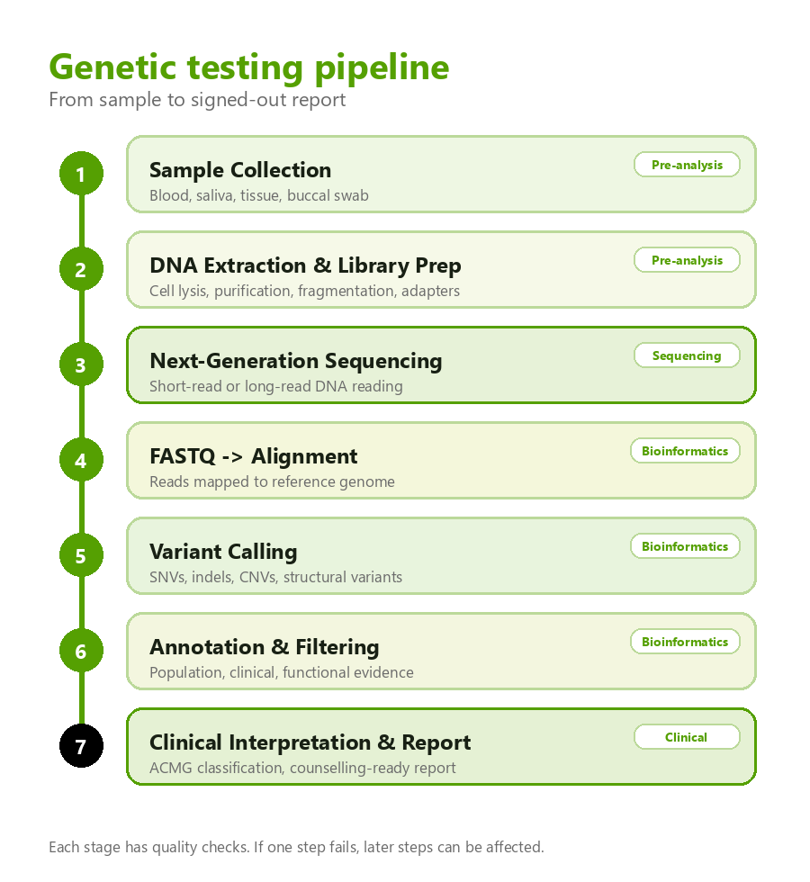
Within this, the first step is alignment. This means each small DNA read is matched to its likely place in a standard human DNA map, called the reference genome. If the raw data is like a pile of loose sentences, alignment is the step that places those sentences back on the correct pages.
After alignment, another software step looks for differences between the patient’s DNA and the reference map. These differences are called variants. The main types include:
- Single nucleotide variants (SNVs): one DNA letter is changed to another. For example, a C may become a T.
- Insertions and deletions (indels): small pieces of DNA are added or missing.
- Copy number variants (CNVs): a person has too many or too few copies of a DNA section.
- Structural variants (SVs): larger pieces of DNA are moved, flipped, deleted, or duplicated.
A whole-genome test can find millions of variants in one person. The hard part is not finding differences. The hard part is deciding which differences, if any, explain the patient’s condition.
To make the list manageable, the lab compares variants with trusted databases and scientific evidence. For example, a variant that is common in healthy people is unlikely to explain a rare disease. A variant in a gene already linked to the patient’s symptoms deserves closer review. These filters can reduce millions of variants to a smaller list for expert interpretation.
Clinical Interpretation: The Hardest Stage
Stage 5: Variant Classification: The Science and Its Limits
Sequencing is the technology. Interpretation is the judgement. The filtered list of variants does not arrive pre-labelled as disease-causing or harmless. A trained analyst or clinical geneticist must weigh the evidence to label them as such.
To keep this process consistent, genetics professionals use international guidelines. The most widely used system comes from the American College of Medical Genetics and Genomics and the Association for Molecular Pathology. These guidelines place variants into five categories.
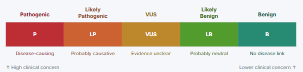
Classification is based on different types of evidence, including:
- Population data: Is the variant common in healthy people? If it is very common, it is unlikely to cause a rare disease.
- Functional data: Do laboratory studies show that the variant damages the gene or protein?
- Computer predictions: Do software tools predict that the change is likely to disrupt normal function? These tools can help, but they are not enough on their own.
- Family pattern: Does the variant appear in relatives who have the condition and stay absent in relatives who do not?
- Disease mechanism: Does the gene normally cause disease in this way, and does the patient’s condition match what is known about that gene?
The VUS Problem and Why It Matters
A Variant of Uncertain Significance, or VUS, means a DNA change was found but there is not enough evidence to say whether it causes disease. This is common in genetic testing. For families who have waited years for an answer, a VUS can be frustrating because it is neither a clear diagnosis nor a clear dismissal.
A VUS usually represents our current knowledge gap, not a failed test. The variant may be too rare, too new, or not well studied. More evidence may come from family testing, published patient reports, laboratory studies, better databases, or AI tools that help researchers collect evidence faster. Until then, a VUS should usually not be treated as a confirmed diagnosis.
Reading the Report
Stage 6: The Genetic Report
The genetic report is the final output of the testing process. It should turn complex laboratory and interpretation work into a clear medical statement. A good report includes:
- Patient and test information: who was tested, why the test was ordered, what sample was used, and what method was used.
- Result summary: a short statement of the main finding in plain language, followed by the technical details needed for the medical record.
- Variant details: the gene, the exact DNA change, whether one or both gene copies are affected, and the classification of the variant.
- Interpretation: what the result means for the patient, whether it explains the symptoms, how it fits the family pattern, and what follow-up is recommended.
- Secondary or incidental findings: some broad tests may also find important health risks unrelated to the reason for testing. These are reported only under defined clinical policies.
- Recommendations: next steps such as confirmatory testing, testing relatives, specialist referral, medication guidance, or research options for unresolved cases.
Genetic results should be explained with genetic counselling whenever possible. The information is personal and can affect relatives as well as the patient. A result without enough explanation can create confusion or anxiety, even when the result is technically correct.
Choosing the Right Test
Not every question needs the biggest test. The right choice depends on the symptoms, the suspected condition, the type of genetic change being considered, cost, and turnaround time.
| Test Type | What It Analyses | Cost | Turnaround | Best Suited For |
|---|---|---|---|---|
| Targeted Gene Panel | A selected set of genes linked to a specific condition or group of conditions | $ | 2-4 weeks | When symptoms point strongly to a known condition, such as hereditary breast cancer or cardiomyopathy |
| Whole Exome Sequencing (WES) | Most protein-coding genes, which make up about 1-2% of the genome | $$ | 4-6 weeks | Undiagnosed rare disease or complex symptoms, especially when a targeted panel is negative |
| Whole Genome Sequencing (WGS) | Nearly all DNA, including regions outside genes | $$$ | 4-8 weeks; rapid testing may be faster for critically ill newborns | Long diagnostic odysseys, suspected large DNA changes, or urgent neonatal cases |
| RNA Sequencing (RNA-seq) | How genes are being read and used in a tissue | $$ | 2-4 weeks | When DNA testing is unclear, especially if a splicing problem is suspected |
| Chromosomal Microarray (CMA) | Larger missing or extra DNA sections across the genome | $ | 2-3 weeks | Developmental delay, congenital anomalies, or suspected chromosome-level changes |
For many undiagnosed rare disease cases, whole genome sequencing is becoming an important option, especially when the child and both parents are tested together. This family-based approach can show whether a variant is inherited or newly appeared in the child, which can make interpretation much stronger.
AI, Multi-Omics, and What Comes Next
Genomics can now generate data faster than experts can interpret it. The next major improvements will come not from better sequencing machines, but from better ways to understand the results and connect them to real patient care.
Artificial Intelligence and Variant Prioritisation
AI tools can help rank variants by how likely they are to matter. These tools may look at many clues at once, such as whether the affected gene is important, whether the changed part is conserved across species, and whether the patient’s symptoms match known gene-disease patterns.
For clinical use, AI must show the evidence behind its suggestions. A score alone is not enough. Doctors and laboratory teams need to know why a variant was prioritised. AI should help experts work faster, not replace expert judgement.
Multi-Omics Integration
DNA testing is powerful, but it is only one layer of biology. Sometimes RNA testing can show whether a DNA change disrupts how a gene message is made. Protein testing can show whether the protein is missing or reduced. Metabolite testing can show downstream effects in the body. Combining these layers can help solve cases that DNA alone cannot explain.
Long-Read Sequencing and the Diagnostic Gap
Short-read sequencing has blind spots. It can miss some repeated sequences, complex rearrangements, or changes in difficult regions of DNA. Long-read sequencing can read longer stretches at once, which helps uncover some of these hidden changes. Early studies suggest it can add diagnoses for some patients whose standard testing was negative.
The Democratisation of Genomics
The next decade will also be shaped by who has access to genetic testing and whose data is represented in genetic databases. India is an important example because of our large population and the number of families living with undiagnosed rare diseases. Genomic medicine cannot simply import all its evidence from Europe and North America. It needs local expertise, local data, and systems that protect patient privacy while allowing useful interpretation. This matters scientifically too because when South Asian, African, and Latin American populations are under-represented in databases, patients from those groups are more likely to receive uncertain results.
Conclusion
Genetic testing is powerful, but it is also a careful chain of steps. A useful result depends on sample quality, sequencing quality, software analysis, reference databases, and expert interpretation. Weakness at any step can weaken the final answer.
The field has changed quickly. Sequencing is faster and cheaper than it was a decade ago. Software can process data in hours rather than months. Databases now contain far more information about genes and variants than before.
Still, genetic testing has limits. Many variants remain uncertain. Even the most complete test may not find an answer for every family. Some changes are hard to detect, some genes are not well understood, and some results depend on evidence that has not yet been collected.
These limits are not fixed forever. Every new patient study, family study, database update, and laboratory experiment helps the field improve. More diverse datasets will be especially important because they can reduce uncertainty for populations that have been under-represented in genetic research.
The practical takeaway is simple. A genetic test is not just a yes-or-no result. It is a structured medical interpretation of DNA. When done well, it can give families a name for what is happening, guide care, and show what steps may come next.
References
References are organised by the order in which the relevant concepts appear in the article.
- Molster CM, Urwin D, Di Pietro L, et al. Survey of healthcare experiences of Australian adults living with rare diseases. Orphanet J Rare Dis. 2016;11:30. doi:10.1186/s13023-016-0409-z.
- Wetterstrand KA. DNA Sequencing Costs: Data from the NHGRI Genome Sequencing Program. Available at: genome.gov/sequencingcostsdata. Accessed May 2026.
- Collins FS, Green ED, Guttmacher AE, Guyer MS. A vision for the future of genomics research. Nature. 2003;422(6934):835-847. doi:10.1038/nature01626.
- Karczewski KJ, Francioli LC, Tiao G, et al. The mutational constraint spectrum quantified from variation in 141,456 humans. Nature. 2020;581(7809):434-443. doi:10.1038/s41586-020-2308-7.
- Sanger F, Nicklen S, Coulson AR. DNA sequencing with chain-terminating inhibitors. Proc Natl Acad Sci USA. 1977;74(12):5463-5467. doi:10.1073/pnas.74.12.5463.
- Bentley DR, Balasubramanian S, Swerdlow HP, et al. Accurate whole human genome sequencing using reversible terminator chemistry. Nature. 2008;456(7218):53-59. doi:10.1038/nature07517.
- Jain M, Olsen HE, Paten B, Akeson M. The Oxford Nanopore MinION: delivery of nanopore sequencing to the genomics community. Genome Biol. 2016;17(1):239. doi:10.1186/s13059-016-1103-0.
- Vasimuddin Md, Misra S, Li H, Aluru S. Efficient architecture-aware acceleration of BWA-MEM for multicore systems. IEEE IPDPS. 2019:314-324. doi:10.1109/IPDPS.2019.00041.
- Schneider VA, Graves-Lindsay T, Howe K, et al. Evaluation of GRCh38 and de novo haploid genome assemblies. Genome Res. 2017;27(5):849-864. doi:10.1101/gr.213611.116.
- Landrum MJ, Lee JM, Benson M, et al. ClinVar: improving access to variant interpretations and supporting evidence. Nucleic Acids Res. 2018;46(D1):D1062-D1067. doi:10.1093/nar/gkx1153.
- Amberger JS, Bocchini CA, Scott AF, Hamosh A. OMIM.org: leveraging knowledge across phenotype-gene relationships. Nucleic Acids Res. 2019;47(D1):D1038-D1043. doi:10.1093/nar/gky1151.
- Rentzsch P, Witten D, Cooper GM, Shendure J, Kircher M. CADD: predicting the deleteriousness of variants throughout the human genome. Nucleic Acids Res. 2019;47(D1):D886-D894. doi:10.1093/nar/gky1016.
- Ioannidis NM, Rothstein JH, Pejaver V, et al. REVEL: an ensemble method for predicting the pathogenicity of rare missense variants. Am J Hum Genet. 2016;99(4):877-885. doi:10.1016/j.ajhg.2016.08.016.
- Jaganathan K, Kyriazopoulou Panagiotopoulou S, McRae JF, et al. Predicting splicing from primary sequence with deep learning. Cell. 2019;176(3):535-548.e24. doi:10.1016/j.cell.2018.12.015.
- Richards S, Aziz N, Bale S, et al. Standards and guidelines for the interpretation of sequence variants: ACMG/AMP joint consensus. Genet Med. 2015;17(5):405-424. doi:10.1038/gim.2015.30.
- Stenson PD, Mort M, Ball EV, et al. The Human Gene Mutation Database: towards a comprehensive repository. Hum Genet. 2017;136(6):665-677. doi:10.1007/s00439-017-1779-6.
- Miller DT, Lee K, Chung WK, et al. ACMG SF v3.0: 73 genes recommended for return of secondary findings. Genet Med. 2022;24(7):1407-1414. doi:10.1016/j.gim.2022.04.006.
- Yang Y, Muzny DM, Reid JG, et al. Clinical whole-exome sequencing for the diagnosis of mendelian disorders. N Engl J Med. 2013;369(16):1502-1511. doi:10.1056/NEJMoa1306555.
- The 100,000 Genomes Project Pilot Investigators. 100,000 Genomes Pilot on rare-disease diagnosis preliminary report. N Engl J Med. 2021;385(19):1868-1880. doi:10.1056/NEJMoa2035790.
- Cheng J, Novati G, Pan J, et al. Accurate proteome-wide missense variant effect prediction with AlphaMissense. Science. 2023;381(6664):eadg7492. doi:10.1126/science.adg7492.
- Mantere T, Kersten S, Hoischen A. Long-read sequencing emerging in medical genetics. Front Genet. 2019;10:426. doi:10.3389/fgene.2019.00426.
- Del Gobbo GF, Boycott KM. The additional diagnostic yield of long-read sequencing in undiagnosed rare diseases. Genome Res. 2025;35(4):559-571. doi:10.1101/gr.279970.124.
- Eisfeldt J, Ek M, Nordenskjold M, Lindstrand A. Toward clinical long-read genome sequencing for rare diseases. Nat Genet. 2025;57(6):1334-1343. doi:10.1038/s41588-025-02160-y.
- Slavin TP, Van Tongeren LR, Behrendt CE, et al. Prospective study of cancer genetic variants: variation in reclassification rate by ancestry. J Natl Cancer Inst. 2018;110(10):1059-1066. doi:10.1093/jnci/djy021.
- Nguengang Wakap S, Lambert DM, Olry A, et al. Estimating cumulative point prevalence of rare diseases: analysis of the Orphanet database. Eur J Hum Genet. 2020;28(2):165-173. doi:10.1038/s41431-019-0508-0.
- Ministry of Health and Family Welfare, Government of India. National Policy for Rare Diseases 2021. New Delhi: MoHFW; 2021. Available at: main.mohfw.gov.in.
- Marwaha S, Knowles JW, Ashley EA. A guide for the diagnosis of rare and undiagnosed disease: beyond the exome. Genome Med. 2022;14(1):23. doi:10.1186/s13073-022-01026-w.
- Stranneheim H, Lagerstedt-Robinson K, Magnusson M, et al. Integration of whole genome sequencing into a healthcare setting: high diagnostic rates in 3,219 rare disease patients. Genome Med. 2021;13(1):40. doi:10.1186/s13073-021-00855-5.